Category

Uncategorized

“Seamus” and the Linear foreign body

By Uncategorized

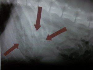
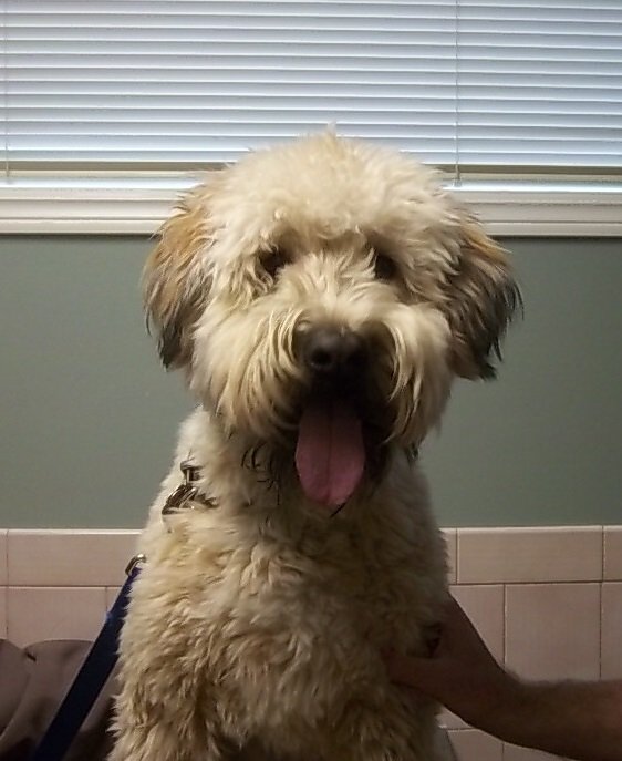
“Seamus” is a usually very energetic, happy 1 year old soft coated wheaten terrier.  He presented to Welland Animal Hospital several weeks ago with a history of lethargy, poor appetite and persistent vomiting.  He was very quiet on presentation, mildly dehydrated, and had a tense and uncomfortable abdomen on palpation.  He had been noted to have chewed some carpet a few days previously.  He was admitted to the hospital for abdominal radiographs which revealed a large multi-focal density in the stomach as well as the small intestine. 


Since Seamus had not eaten in the last 24 hours and had been profusely vomiting we would expect his stomach to be empty.  However, his  stomach was full  suggesting  that a foreign body (likely carpet) was present and was lodged in the stomach and potentially the small intestine. A linear foreign body arises when linear material (carpet strands, rope, thread, ribbon, plastic, cloth, sacks, cassette tapes..) is anchored in one spot while the intestine attempts to move the material towards the anus with normal peristaltic muscular contractions.  Because the material is stuck and immovable (usually in pylorus of stomach-narrow exit to small intestine, or under the tongue in cats with string) the constant movement of the intestine will result in the bunching of the intestine and a partial or complete intestinal obstruction.  Continued peristalsis will cause the foreign material to become taut and eventually to cut into the intestine at the mesenteric border causing leakage at multiple sites and a life threatening infection (peritonitis).  Multiple perforations of bowel are associated with high levels of mortality.

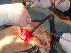
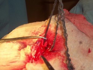
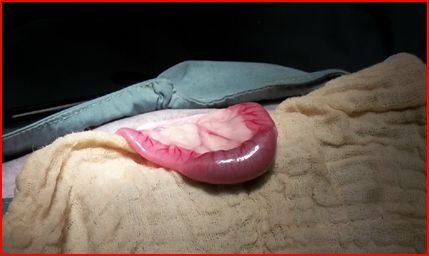
An exploratory abdominal surgery was recommended and Seamus was admitted to the hospital immediately, started on IV fluids, antibiotics and stomach protectants.  A routine ventral abdominal exploratory revealed a large distended stomach and a linear foreign body extending from the stomach to the proximal -mid jejunum (small intestine). The foreign material carpet was removed through an incision in the stomach (gastrotomy)- see below

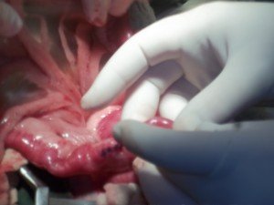
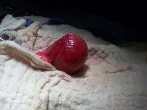
  and two incisions into the small intestine (enterotomies)- see above.  The intestine all appeared viable with no leakage or vascular compromise.  The pancreas was mildly red and swollen. All surgical sites were closed routinely and the abdomen flushed with saline ( below see one of the sutured enterotomy sites).

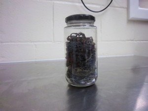
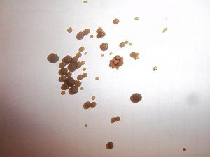
 Routine closure of the adominal wall was performed and Seamus’s recovery was uneventful. The carpet was collected in a jar so the owner’s could visualize without the associated smell.

Seamus was started on some antiemetics ( to control vomiting) and medication to encourage intestinal motility, as well as continued supportive care with IV fluids and antibiotics.  His appetite gradually returned, the vomiting stopped and his vibrant personality gradually returned.  He was discharged to his caring owners who did an excellent job of nursing him along for the next few days as his appetite and bowel movements normalized.  We are happy to report that Seamus has been back for his final recheck and is doing great.  He gave us all an outstanding greeting and enjoyed a few well earned treats.

Welland Animal Hospital Case of the Month – “Moey” Difficulty urinating

By Uncategorized

“Moey” is a very sweet 9 year old male cat who recently presented to Welland Animal Hospital with a history of weakness, lethargy, poor appetite, breathing difficulties – panting  and no urine or BM’s noted recently. Physical examination revealed moderate dehydration, painful abdomen on palpation with a large turgid unexpressable bladder.  Respiration’s were shallow and increased with palpation of bladder, but no other abnormalities were auscultated.  A preliminary diagnosis of urinary obstruction was made and Moey was admitted to the hospital for blood-work, intravenous fluids, and urinary catheter placement to unobstruct the urethra as well as continued monitoring. 

Urinary obstruction is a common problem most usually affecting younger male cats.  Most commonly the cause is a large number of crystals/stones  in the urine which block the narrow male urethra making it impossible to urinate.  This is a life threatening condition as electrolyte and other metabolic changes will eventually result in cardiac arrhythmias and death.  Other causes of obstruction may be tumours of the bladder/urethra, blood clots after trauma (hit by car) urethral mucus plugs…There are numerous factors that may predispose some male cats to form crystals/stones that lead to urinary obstruction including, diet, genetics,  size of urethra, water consumption and concentration of urine to name a few.  Any cat showing signs of straining to urinate, blood in urine, vocalizing during urination, urinating frequently, not eating, vomiting, painful abdomen should be evaluated by a veterinarian immediately.

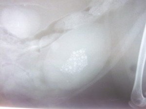
Moey was started on IV fluids immediately to correct dehydration and electrolyte abnormalities and promote urine production. Blood work revealed severe azotemia (elevated kidney values BUN+CRT) as well as severely elevated phosphorous and potassium secondary to being unable to urinate, and dehydration.  Primary kidney disease could  not be ruled out at this time but it was hoped that the azotemia was due solely to post renal obstruction and would not be permanent.  Moey was sedated and given pain medication and his urinary obstruction relieved through flushing of the urethra multiple times.  A urinalysis showed marked hematuria (red blood cells in the urine) and a small number of crystals. A silicone urinary catheter with a closed urinary collection system was placed for 72 hours.  The bladder needed to be kept small for a period of time so the muscular layer  of the bladder wall could recover from the serious stretching that occurs during obstruction. Repeat blood work after 48 hours of IV fluids and unobstruction revealed normal electrolyte and kidney values and Moey was feeling much better.  However, after the urinary catheter was removed Moey once again had difficulty urinating and had  a poor urine stream. Radiographs of his abdomen were taken and revealed a bladder full of small radio-dense stones.  Because of the likelihood of re-blocking and an already compromised urinary system a cystotomy was recommended over any attempt to dissolve the stones with a special diet.


A routine ventral cystotomy (surgically opening the bladder to remove the stones) was performed and all stones removed after multiple flushes through a urinary catheter.

The bladder wall was extremely thickened secondary to chronic inflammation but the surgery was successful and all stones were removed as seen on the post -op radiograph. The end of the urinary catheter can also be visualized in the radiograph.

Moey’s urinary catheter was kept in place for a few days and he was discharged from the hospital urinating well.  He continues to do well at home on a special diet while we wait for the bladder stone analysis from the lab after which we can formulate a plan to attempt to prevent the recurrence of the stones through diet, medications ..etc.


We recommend  radiographs every 4-6 months initially to check for stones and monitor his kidney values and urine for any signs of disease/recurrence. Unfortunately some cats will continue to form stones despite dietary and medical management  and may require additional surgeries in the future.

The timely presentation of Moey to Welland Animal Hospital  by his loving owners was paramount in a successful outcome.  Moey was a pleasure to work with and we wish him all the best in the years to come.


Canine and Feline Cognitive Dysfunction

By Uncategorized

Canine and feline cognitive dysfunction (dementia – similar to Alzheimer’s in people) is usually diagnosed in dogs over 10 years of age and cats over 12 years.  It is a disease recognized by an onset of behaviour changes in a geriatric pet which cannot be attributed to any other medical conditions.  It is a gradually progressive disease in which older animals show more clinical signs.

Dogs with cognitive dysfunction display signs in five categories:

Disorientation – stares into space at ceiling or wall, barks at the ceiling, gets stuck in corners, wanders aimlessly, confused, doesn’t recognize family members or commands

Interactions – apathetic, indifferent, doesn’t participate in activities they used to enjoy

Sleep/wake cycle – keeps owner up at night and sleeps all day, howls or barks at night, sleeps more in a 24 hour period

House Training – Walks outside and seems confused, then urinates or defecates inside, no longer signals to go outside

Activity/anxiety/aggression – plays less, increased anxiety or aggression

Cats with cognitive dysfunction generally show signs associated with:

House Soiling, excessive vocalization – especially at night, apathy, less play and change in aggression.

If your older pet is experiencing any of these signs it is essential that your veterinarian is consulted to rule-out any underlying medical condition which may be an underlying cause (ex. arthritis, diabetes, kidney disease, seizures, cancer, hearing or vision problems….)  In most cases a thorough physical exam and some diagnostic tests (blood work, urinalysis, radiographs..) can rule out a medical problem so that treatment for cognitive dysfunction can begin.

Treatment for cognitive dysfunction is similar to other behavioural problems and includes behaviour modification, enrichment, safety recommendations and medication as needed.  In dogs, Anipryl (selegiline)  is an approved drug for cognitive dysfunction and can help restore the sleep/wake cycle and slow the progression of the disease.  Nutritional therapy and some nutraceuticals (SAME) have been shown to be helpful in treatment of cognitive dysfunction in cats and dogs with limited to no side effects.  Environmental enrichment tools (ex. variety of toys..), predictable routines, retraining of certain behaviours (house training) are very important in the success of any treatment plan.  In geriatric cats that are no longer using the litter box, once medical issues have been ruled out, the height, size and location of the litter box should be evaluated.  Often times the older cat may have difficulty with stairs, and climbing in and out of the litter box, so having one available on all floors that is large and easy to get into ( low sides) is very important.

As our pets live longer, geriatric behavioural/medical issues will become more common. Regular consultation with your veterinarian is paramount in diagnosing and treating these issues and improving the quality of your old friends golden years.

Reference – lecture OVMA conference Toronto Jan 2013 – Dr. Lisa Radosta (DACVB) Florida Veterinary Behaviour Service


Hit By Car – Degloving Wound “Bella”

By Uncategorized

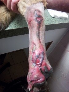
“Bella” is a 4 year old retriever mix who was hit by a car Nov 1, 2012 and was dragged for a short distance.  She was non-weight bearing on her left rear leg with a significant degloving wound.  Her left hock was swollen but appeared stable and there were no palpable fractures.  Although in obvious discomfort she was otherwise stable.  Degloving injuries are commonly associated with motor vehicle trauma and occur when the body is dragged along a hard coarse abrasive surface (asphalt) which literally removes the soft tissue (skin, muscle) and sometimes the ligaments, bone, and nails as well.  Usually the wounds are infected as dirt and other debris are driven into the wound.  Large degloving wounds on the extremities are always a challenge due to the lack of skin and  difficulty closing wounds surgically.  Some wounds will heal secondarily with multiple bandage changes while others may require, delayed surgical closure +/- use of skin grafts, pedicle grafts, or wound care products that support and maintain a healing environment.(ACell Vet)


“Bella” was given pain medication and sedated so the wounds could be clipped, cleaned, debrided and flushed extensively.  Antibiotics were started, and a soft padded dressing was placed over an application of granulated sugar on the wound surface.  Sugar dressings have been used successfully for many years  and effectively clean the wound.  The sugar creates an environment in which bacteria cannot grow but healthy granulation tissue can form.  It is an inexpensive wound care ingredient and it is relatively easy to do bandage changes as it is relatively non-adherent. Bandage changes were done daily – initially with sedation and pain control.  The owner’s were able to do a lot of the bandage changes and the wound was closely monitored over the next 2 weeks with remarkable results.  Weight bearing gradually improved as the wound and the swelling improved.


Once healthy granulation tissue was present and minimal discharge was observed, the sugar was discontinued and and a non aderent dressing placed every other day.  The owner’s were extremely diligent in keeping the bandage clean and dry and changing it if it was soiled.  This was instrumental in the healing of this degloving wound. This is the wound 4 & 6 weeks after the trauma  completely healed.


Welland Animal Hospital Case of the Month – “POE” and a Noodle

By Uncategorized


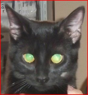
“Poe” is a very cute 1.5 year old black male cat who came to Welland Animal Hospital with a history of multiple episodes of severe vomiting, no  interest in food and acting very quietly over the last 12 hours.  He had no previous medical problems except for some vomiting 1 month previously after chewing on a flotation foam “pool noodle”.  Bloodwork was unremarkable, and abdominal radiographs revealed no radio-dense foreign body but there was an unusual gas pocket in the small intestine.  Abdominal palpation was unremarkable except for some mild discomfort.  “Poe” was placed on IV fluids to maintain his hydration, and medications to control his vomiting and discomfort.  This initial supportive care did not seem to alleviate his  abdominal problems and repeat abdominal radiographs revealed a similar gas pocket and slight dilation of a small portion of his small intestine.  An exploratory surgery was recommended in an attempt to discover what was causing “Poe”‘s problems.

A thorough exam of his abdomen revealed an inflamed pancreas and duodenum (first part of the small intestine) – consistent with pancreatitis and possible local irritation from a foreign body.


Further along his small intestine  (mid jejunum)  a foreign body was palpable which completely blocked the passage of gas and fluid.

The intestines had lost there normal motility (ileus).  The intestine in which the foreign body was found looked  viable and an incision was made just beyond the obstruction (enterotomy) and the foreign material was gently pushed out of the incision.

The foreign material was – you guessed it – a large piece of compressed foam from the previous chewed “pool noodle” – which had probably sat in the stomach for a month before proceeding into the small intestine and causing an obstruction.  The intestinal incision was sutured and the abdominal incision routinely closed.  “Poe” recovered uneventfully and is doing very well.  No more “noodles” for you.

Bee, wasp, hornet and yellow jacket stings and your dog

By Uncategorized

These insects inject their venom when the unsuspecting dog either steps on them or disturbs or frightens them.  Bees lose their barbed stinger after stinging and die where wasps, hornets and yellow jackets may sting many times.  Most dogs that are stung will suffer from mild local reactions which may include redness, pain, and local swelling at the sting site.  Diagnosis is based on the observation of the sting and the appropriate clinical signs. Treatment may include cool compresses, antihistamines; corticosteroid lotions… consult your veterinarian.  Rarely a sting may cause a regional reaction (swelling, rash) which may require systemic antihistamines and anti-inflammatory.  Very, very rarely a severe life threatening anaphylactic reaction (collapse, severe hypotension, breathing difficulty, pale mucous membranes…) may be caused within 10 minutes of the sting which requires immediate veterinary intervention.  Treatment for anaphylaxis will include IV fluids, antihistamines, anti-inflammatory, and epinephrine.   Owners of dogs with a known history of anaphylaxis to insect bites should set up a bee sting kit with their veterinarian’s guidance which would include an epi pen of suitable size for their pet.  Immediate veterinary intervention should always be sought in the case of anaphylaxis; however the kit may enable a dog in crisis to survive especially if veterinary assistance is not locally available. (Camping, after hours…)

Thank-you for your support in the ride to conquer cancer

By Uncategorized

Dr. Fidler would like to thank all those clients, friends and family who supported him in the ride to conquer cancer on Jun 9-10, benifitting the Princess Margaret – one of the top 5 cancer research hospitals worldwide.  The ride raised over 18 million dollars making it the largest single cycling fund raiser in the world.  This is the third year that he has participated and once again it was a very rewarding and awe inspiring experience.