All Posts By

wellandah

Cutaneous Mast Cell Tumors in Dogs

By Uncategorized

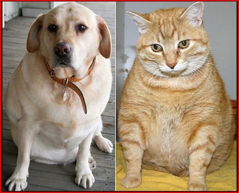
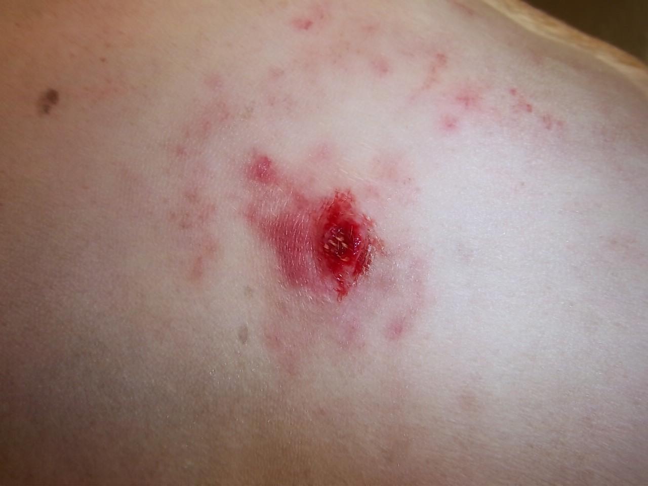
Cutaneous  mast cell tumors are the most common malignant skin tumor in dogs.  The most common finding on presentation is a lump which may be of varying size, consistency and is sometimes ulcerated or very inflamed and itchy. The median age at diagnosis is 8-9 years but dogs of any age can be affected.  Certain breeds are much more commonly affected such as the Boxer, pug, Boston terrier, Labrador retriever, golden retriever, beagle and bull terrier.  Mast cells originate in the bone marrow and are eventually found in all body tissues but are most concentrated in the skin, respiratory tract and intestinal tract.  These cells have a wide array of normal functions in the tissues and it is currently not known why in some individuals they begin to proliferate out of control (mast cell tumor).

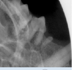
If a skin mass is discovered on a dog the initial step in diagnosis will be a FNA (fine needle aspirate).  This involves placing a small needle into the mass and getting a small sample of cells to allow for diagnosis and presurgical planning.  Most MCT can be identified in this manner.  (see below a FNA of a grade 2 MCT – note the dark granules within the cells characteristic of a MCT)


Some grades of these tumors are very diffuse and not well demarcated from normal tissue so that very wide surgical margins are required for there complete excision.  This can be difficult depending on the body location.  After the mass has been removed the lump is submitted to a pathologist for grading and to assess the margins.

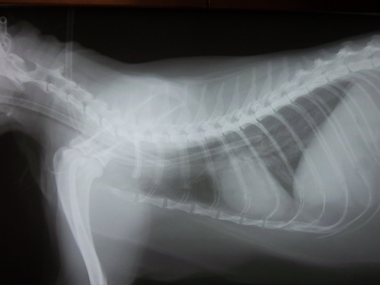
These type of tumors are graded from 1-3.  Grade 1 tumors are well differentiated and confined to the skin. Complete excision is usually curative. Grade 2 are intermediate and are usually subclassified as low – intermediate grade vs intermediate – high grade, 65 % are cured surgically but recurrence locally and metastasis (tumor spread are possible). Grade 3 tumors are the least favorable with local recurrence and metastasis likely and are almost always fatal. Clinical staging tests may establish that distant spread has occurred (radiographs, ultrasound, bloodwork…)

The most important treatment is wide margin resection of the mast cell tumor.   Grade 2 intermediate and grade 3 tumors or those tumors that reoccur will benefit from radiation therapy and chemotherapy in addition to consultation with an oncologist/surgeon.

After successful surgical removal of the mass careful monitoring of the surgical site is required. No recurrence after 1 year is encouraging but a cure is always a guarded term.

Skin masses in dogs (especially in susceptible breeds) should be evaluated promptly by your veterinarian and removed with wide surgical margins if a MCT is suspected.  Post surgical grading of the tumor will determine follow up treatment and likely prognosis.


“Precious” – Chronic Gingivostomatitis

By Uncategorized

“Precious” is an 8 year old female spayed cat who was referred to Welland Animal Hospital for a condition of severe chronic inflammation of the oral cavity.  This inflammation extends beyond the gingiva to the mucous lining of many structures in the mouth – a disease known as chronic gingivostomatitis. (this picture does not due justice to  the extent of the severe inflammation – especially in the back of the mouth).

001

This severe inflammation results in oral pain and discomfort and cats will eventually stop eating.  The cause of this condition is unknown but is most likely an immune mediated reaction secondary to bacteria (plaque) vs food antigens vs environmental antigens vs viruses vs other.

Bloodwork (CBC + biochemistry) on Precious was within normal limits and she was negative for feline leukemia and feline immunodefiency virus.  Routine dentistries and homecare had not controlled the inflammation in the oral cavity and despite pain medication Precious had stopped eating for several days prior to presentation.  In most cases the objective for these cats is to obtain a pain free oral cavity which is best achieved by doing a full mouth extraction (removal of all the teeth).  Domestic cats do not need teeth to live long and happy lives and this provides the best long term resolution of inflammation – about 60 % complete remission.  Some cats may still require adjunctive treatment post full mouth extractions.  Referral to a veterinary dentist was advised but was not possible in this case so Precious was referred to our hospital.

Whole mouth intra oral radiographs were taken (see one below)

Capture

local anesthetics were given ( to aid in post -operative pain control) and all teeth and roots were extracted and alveoli were cleaned of all periodontal ligaments. Post operative rads revealed all teeth were completely removed.  All extraction sites were closed with gingival flaps using absorbable sutures.

005

Because of Precious’s poor appetite prior to the dental an esphagotomy tube was placed in her left neck during the dental so she could be fed post operatively. (see radiograph of chest showing feeding tube placement)

X-Ray

Precious recovered well from the long aneasthetic and feedings were begun the next day through the esophagotomy tube.  She was gradually weaned off IV fluids and pain medications and began eating  some on her own 2 days post op.  The feeding tube was removed 4 days post – op and Precious was sent home.  She was subsequently treated with anti-inflammatories 2 weeks post dental which seem to have greatly reduced the inflammation in the caudal oral cavity and she is currently eating well.

Chronic gingivostomatitis is a painful debilitating disease which at this time is best treated by whole mouth extraction.  This is the best hope for a pain free mouth in cats that are suffering from this particular disease.  Routine oral  and dental examination by your veterinarian is essential in diagnosing this condition early.


brown_ white puppy on grasses

Heart Failure in small breed dogs

By Uncategorized

Heart disease is one of the most common diseases that affect small breed dogs that we see at the Welland Animal Hospital (ex Chihuahuas, Toy poodles, Cavalier King Charles spaniels, Pekinese, Shih Tzu,….)  Some estimates suggest 1/3 of small dogs over the age of 10 may be affected with heart disease caused by mitral valve disease.  The mitral valve is located between the left atrium and left ventricle and when functioning normally forms a perfect seal preventing blood from flowing backwards (into the left atrium) as the ventricle contracts and pushes blood forward to the rest of the body.  When this valve is defective some blood will leak backwards which eventually leads to heart enlargement and decreased blood being pumped in a  forward direction.

Most dogs with mitral valve disease will initially have few if any clinical signs.  Initially a heart murmur detected by your veterinarian during a routine physical exam may be the only sign of mitral valve disease.  A murmur is the result of turbulent blood flow which occurs across the faulty valve and is easily detected with a stethoscope during a routine physical examination.  Heart murmurs are graded from 1-6 depending on their severity.  If a heart murmur is detected your veterinarian may suggest one or  more of the following depending on the number of clinical signs noted 1) frequent physical exams to monitor your pet 2) chest radiographs to determine heart size (baseline) and appearance of lungs (signs of respiratory disease – bronchitis  vs  pulmonary edema ….), 3) bloodwork to assess for other diseases (kidney function, liver disease, heartworm disease…), 4) an EKG (electrocardiogram) – if an abnormal rhythm is heard, and 5) cardiac BNP (blood test which becomes elevated as the heart enlarges) 6) ultrasound of the heart – useful if the diagnoses is complicated, can visualize the abnormal flow across the valves and measure chamber size.

Below is a lateral chest radiograph of a normal dog

Normal

CHF

Above is a lateral chest radiograph of a dog in heart failure (coughing, difficulty exercising..) with an enlarged heart and evidence of pulmonary edema (base of heart and caudal lung lobes)

Some dogs will continue to show no clinical signs of heart disease for months to many years after a heart murmur is first detected, while other dogs may progress more rapidly.  Common clinical signs associated with progressive heart disease include the following: progressive cough – especially at night, difficulty breathing, difficulty exercising, restless at night – difficulty sleeping, increased heart rate, irregular heart rate, poor appetite, weight loss, and fainting spells – syncope to name a few.

There is to date no cure for heart failure caused by mitral regurgitation but medications can reduce the symptoms and improve the quality of life as well as the life expectancy of most dogs.  Medications frequently used to treat heart failure include diuretics (furosemide) to decrease fluid accumulation in the lungs, vetmedin (pimobendan) to increase strength of heart contraction and dilation of blood vessels, and ACE-inhibitors which improve the blood flow through vessels.

Heart disease is a chronically progressive disease that can be effectively treated for varying lengths of time.  It is essential to give medications as instructed and to recheck with your veterinarian should you notice any change in your dogs clinical signs or as recommended by your veterinarian.  A proper diet lower in sodium is also beneficial as is a proper exercise regime.


black dog with red lash

Welland Animal Hospital – Case of the Month – ‘Shannon’ – Swollen Digit

By Uncategorized

“Shannon” is a very sweet 12 year old female spayed Gordon Setter who presented initially with a swollen left hind medial digit (toe) with an abnormal or deviated nail.  The toe had gradually become more swollen and the nail had been abnormal for several weeks. There was a small amount of discharge noted at the nail base.  There was no known trauma.  The toe was uncomfortable on palpation although Shannon is a very stoic dog. Blood work was unremarkable.  Potential rule outs for a swollen digit include trauma, chronic infection (bacterial, fungal)  or neoplasia (cancer).

Radiographs of Shannon’s LH paw were recommended and can be seen below.  There is a moderate ST swelling around the medial digit of the LH paw with bony lysis evident in P3 ( 3rd phalanx) and the distal part of P2( 2nd phalanx). (red arrow).  Bone invasion of the digit on radiographs in older dogs is commonly seen with neoplasia (cancer). Possible tumors include squamous cell carcinoma, melanoma, soft tissue sarcoma, osteosarcoma and mast cell tumors.  Prognosis very much depends on tumor type (ie. how locally aggressive and the  metastatic potential of a particular tumor – ie the potential of the cancer to spread to other parts of the body).

007


The recommendation to amputate the medial digit at the Metatarsal and P1 junction was made (blue arrow on rads).  This would allow for a definitive diagnosis with histopathology (biopsy) as well as remove any discomfort the abnormal digit was causing.  In case the diagnosis was cancerous the amputation was made at a level which appeared healthy on the radiographs and which would leave a large clean margin. The central 2 digits are the primary weight bearing digits so the removal of the medial digit usually does not cause any significant lameness.

The amputation surgery went very well. The toe was removed, blood vessels ligated and the incision closed.  A soft padded bandage was applied and changed every few days and Shannon was sent home with pain medication.  The site healed nicely and Shannon is walking well.

001

The biopsy came back as a nail bed squamous cell carcinoma with clear margins devoid of any tumor. These types of tumors represent about one third of the digital tumors in dogs.  They generally occur in older dogs and more commonly in large breed dogs with black coats.  They are usually solitary tumors and often cause disfigurement or loss of the nail.  This type of tumor has a low metastatic potential (don’t often spread to other areas of the body) and are often successfully treated by amputation.  We are hopeful that Shannon will continue to do well – she and her parents are fantastic – many thanks for the shortbread cookies.


fat dog fat kitten

Obesity in Dogs and Cats – Body Fat Index for determining optimal body weight

By Uncategorized

Obesity in dogs and cats is one of the most common medical issues that we face on a daily basis at Welland Animal Hospital.  The most common causes are dietary (overfeeding vs high  fat/caloric diets), lack of exercise, genetic factors, and medical issues like hypothyroidism for example.  A thorough physical exam by your veterinarian and blood work can often eliminate medical causes of obesity.

Dogs and cats that are obese or even overweight are much more prone to several general health risks including diabetes mellitus, heart disease, hypertension, respiratory disease – COPD (chronic obstructive pulmonary disease), orthopaedic injury (cruciate disease) and exacerbation of arthritis as well as various cancer links.  The sooner that our pets can return to an optimal body weight the healthier they will be.

Body condition scoring by your veterinarian has historically been the way to assess the ideal body weight of your pet or goal weight.  This is an estimate based on the amount of fat palpable along the spine and laterally along the ribs. This can be quite subjective and often times we underestimate the amount of weight loss.  A newer approach utilizes measurements taken on your cat or dog and a body fat index chart.  Four measurements are taken in the dog(see picture below) and six measurements are taken in the cat which are then entered into a computer algorithm to determine the pet’s optimal  body weight.  This has proven to be the most accurate way to determine the ideal weight of most pets.

bf measures


Once the  goal weight has been determined your  veterinarian will recommend a weight loss plan.  The plan should detail the amount of weight to safely lose weekly and usually requires frequent weigh ins.  This plan may include a new innovation in diet from Hills called Metabolic.  This diet was developed to actually change your pets metabolism by altering or down regulating the genes responsible for transferring fat into individual cells.(nutrigenomics) Large blinded feeding trials have shown how effective this diet is in treating obesity.  It is not a traditional weight loss diet ( low fat, low calorie and high fibre) and can be fed throughout the pets lifetime.  Special treats accompany the diet as well for optimal weight management.  In addition to diet, exercise plays a vital role in any weight loss program.  Gradually increasing the amount of exercise (walking – running) in addition to pain medication if required (arthritis..) in addition to  physical therapy and non weight bearing exercise like swimming may all be part of the program. Indoor cats may require more stimulation with various toys, feeding and litter boxes on different levels of the  house….Some dogs may benefit from a weight loss medication called slenterol to reach there ideal body weight.

In most cases the prognosis for attaining an ideal body weight is good if a weight loss plan is initiated and followed.  The innovation of altering a pets metabolism through diet will be a huge factor in alleviating obesity from our pet population.  Ideally,  this is a problem that is better prevented in the first place. Unsure if your pet is overweight – have your veterinarian assess them and consider body measurements for calculating the total body fat index and ideal body weight.