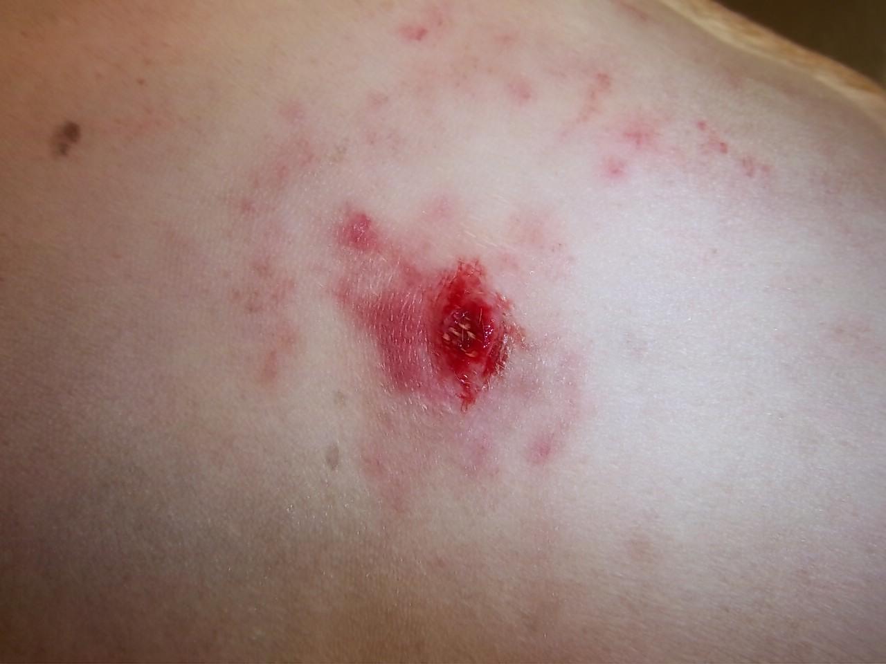
Cutaneous mast cell tumors are the most common malignant skin tumor in dogs. The most common finding on presentation is a lump which may be of varying size, consistency and is sometimes ulcerated or very inflamed and itchy. The median age at diagnosis is 8-9 years but dogs of any age can be affected. Certain breeds are much more commonly affected such as the Boxer, pug, Boston terrier, Labrador retriever, golden retriever, beagle and bull terrier. Mast cells originate in the bone marrow and are eventually found in all body tissues but are most concentrated in the skin, respiratory tract and intestinal tract. These cells have a wide array of normal functions in the tissues and it is currently not known why in some individuals they begin to proliferate out of control (mast cell tumor).
If a skin mass is discovered on a dog the initial step in diagnosis will be a FNA (fine needle aspirate). This involves placing a small needle into the mass and getting a small sample of cells to allow for diagnosis and presurgical planning. Most MCT can be identified in this manner. (see below a FNA of a grade 2 MCT – note the dark granules within the cells characteristic of a MCT)

Some grades of these tumors are very diffuse and not well demarcated from normal tissue so that very wide surgical margins are required for there complete excision. This can be difficult depending on the body location. After the mass has been removed the lump is submitted to a pathologist for grading and to assess the margins.
These type of tumors are graded from 1-3. Grade 1 tumors are well differentiated and confined to the skin. Complete excision is usually curative. Grade 2 are intermediate and are usually subclassified as low – intermediate grade vs intermediate – high grade, 65 % are cured surgically but recurrence locally and metastasis (tumor spread are possible). Grade 3 tumors are the least favorable with local recurrence and metastasis likely and are almost always fatal. Clinical staging tests may establish that distant spread has occurred (radiographs, ultrasound, bloodwork…)
The most important treatment is wide margin resection of the mast cell tumor. Grade 2 intermediate and grade 3 tumors or those tumors that reoccur will benefit from radiation therapy and chemotherapy in addition to consultation with an oncologist/surgeon.
After successful surgical removal of the mass careful monitoring of the surgical site is required. No recurrence after 1 year is encouraging but a cure is always a guarded term.
Skin masses in dogs (especially in susceptible breeds) should be evaluated promptly by your veterinarian and removed with wide surgical margins if a MCT is suspected. Post surgical grading of the tumor will determine follow up treatment and likely prognosis.


